Volume 30, Number 4—April 2024
Dispatch
Isolation of Batborne Neglected Zoonotic Agent Issyk-Kul Virus, Italy
Davide Lelli , Ana Moreno, Sabrina Canziani, Laura Soliani, Maya Carrera, Anna Castelli, Francesca Faccin, Tiziana Trogu, Enrica Sozzi, Gian Luca Cavallari, Matteo Mauri, Fabiana Ferrari, Cristian Salogni, Chiara Garbarino, Chiara Chiapponi, Marco Farioli, and Antonio Lavazza
Figure 1
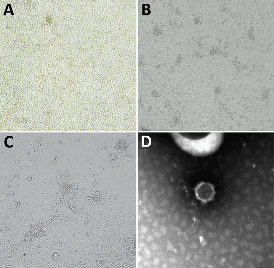
Figure 1. Microscopic appearance of Issyk-Kul virus IT-297348-34/2022, isolated from a Hypsugo savii bat, in study of batborne neglected zoonotic agent Issyk-Kul virus, Italy. A) Issyk-Kul–infected MARC 145 cells, mock infection; original magnification ×10. B) Issyk-Kul–infected MARC 145 cells showing cytopathic effect at 120 hours after infection; original magnification ×4. C) Issyk-Kul–infected MARC 145 cells showing cytopathic effect at 120 hours after infection; original magnification ×10. D) Negative-staining electron microscopy performed on cell supernatants (NaPT 2%), showing a viral particle of 55–60 nm morphologically referable to nairovirus; original magnification ≈x550,000
Page created: March 01, 2024
Page updated: March 20, 2024
Page reviewed: March 20, 2024
The conclusions, findings, and opinions expressed by authors contributing to this journal do not necessarily reflect the official position of the U.S. Department of Health and Human Services, the Public Health Service, the Centers for Disease Control and Prevention, or the authors' affiliated institutions. Use of trade names is for identification only and does not imply endorsement by any of the groups named above.