Skip directly to site content Skip directly to page options Skip directly to A-Z link Skip directly to A-Z link Skip directly to A-Z link
Volume 29, Number 7—July 2023
CME ACTIVITY - Research

Clinical and Mycologic Characteristics of Emerging Mucormycosis Agent Rhizopus homothallicus

Shivaprakash M. Rudramurthy1, Shreya Singh12, Rimjhim Kanaujia, Hansraj Chaudhary3, Valliappan Muthu, Naresh Panda, Abhishek Pandey, Sheetal Thakur, Harsimran Kaur, Anup Ghosh, Ritesh Agarwal, and Arunaloke Chakrabarti4Comments to Author 
Author affiliation: Postgraduate Institute of Medical Education and Research, Chandigarh, India

Main Article

Figure 1

Macroscopic and microscopic characteristics of Rhizopus arrhizus and R. homothallicus isolated from tissue samples from patients enrolled in a 10-month retrospective study, Chandigarh, India, January–October 2021. A, B) Macroscopic appearance of colonies of R. homothallicus (A) and R. arrhizus (B) fungi. C, D) Macro lens image of the colonies showing dark-brown specks in R. homothallicus (C) and black and white dots (salt and pepper appearance) in R. arrhizus (D). E) Photomicrograph from a lactophenol cotton blue mount of R. homothallicus showing multiple reddish brown ornamented zygospores (original magnification ×400). F) Magnified image of E showing unequal suspenser cells and zygospore with prominent spinous projections. G) R. arrhizus showing long, unbranched sporangiophore with nodal rhizoids (original magnification ×200). H) Magnified image of extensively branched rhizoid seen in R. arrhizus. I) Magnified image of sporangiospores of R. arrhizus.

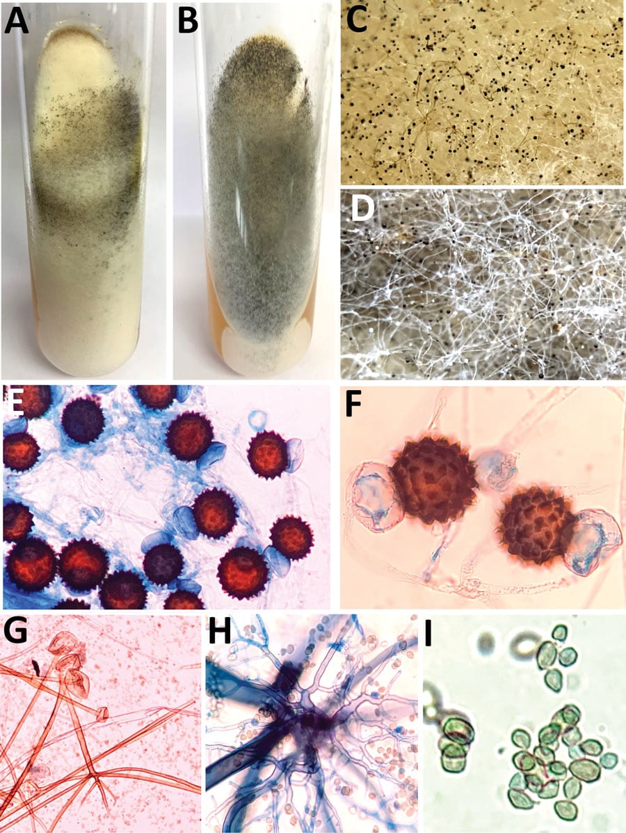
Figure 1. Macroscopic and microscopic characteristics of Rhizopus arrhizus and R. homothallicus isolated from tissue samples from patients enrolled in a 10-month retrospective study, Chandigarh, India, January–October 2021. A, B) Macroscopic appearance of colonies of R. homothallicus (A) and R. arrhizus (B) fungi. C, D) Macro lens image of the colonies showing dark-brown specks in R. homothallicus (C) and black and white dots (salt and pepper appearance) in R. arrhizus (D). E) Photomicrograph from a lactophenol cotton blue mount of R. homothallicus showing multiple reddish brown ornamented zygospores (original magnification ×400). F) Magnified image of E showing unequal suspenser cells and zygospore with prominent spinous projections. G) R. arrhizus showing long, unbranched sporangiophore with nodal rhizoids (original magnification ×200). H) Magnified image of extensively branched rhizoid seen in R. arrhizus. I) Magnified image of sporangiospores of R. arrhizus.

Main Article

1These first authors contributed equally to this article.

2Current affiliation: Dr. B R Ambedkar State Institute of Medical Sciences, Mohali, India.

3Current affiliation: Indian Council of Medical Research, New Delhi, India.

4Current affiliation: Doodhadhari Burfani Hospital, Haridwar, India.

Page created: April 20, 2023
Page updated: June 20, 2023
Page reviewed: June 20, 2023
The conclusions, findings, and opinions expressed by authors contributing to this journal do not necessarily reflect the official position of the U.S. Department of Health and Human Services, the Public Health Service, the Centers for Disease Control and Prevention, or the authors' affiliated institutions. Use of trade names is for identification only and does not imply endorsement by any of the groups named above.
file_external