Purpose
The International Labor Organization (ILO) International Classification of Radiographs of Pneumoconioses is used for epidemiological studies, screening and surveillance of workers exposed to workplace dust, and clinical purposes. The NIOSH B Reader Program certifies physicians in the ILO classification system.
Classification system
The ILO International Classification of Radiographs of Pneumoconioses is a global tool used to:
- Improve worker health surveillance.
- Conduct epidemiological research.
- Make comparisons of statistical data.
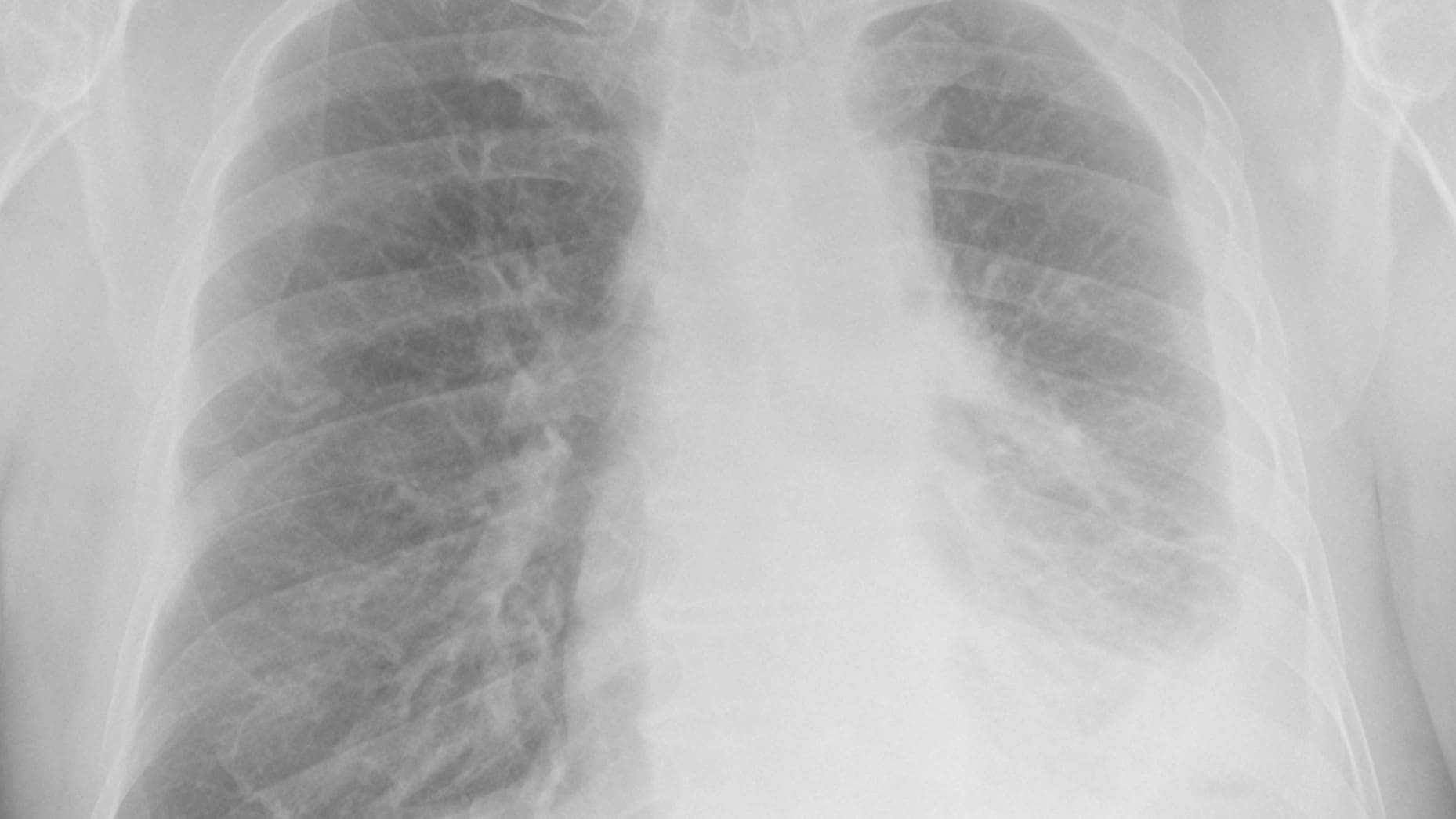
The Classification System includes the Guidelines and two sets of standard images. Standard images represent different types and severity of lung abnormalities. They are used for comparing a person's images during the classification process. The system describes the nature and extent of features associated with different pneumoconioses. They include:
- Coal workers' pneumoconiosis
- Silicosis
- Asbestosis
It deals with:
- Coal workers' pneumoconiosis
- Silicosis
- Asbestosis
Characterizations and terms
In the ILO system, readers must:
- Grade radiograph quality.
- Categorize small opacities according to shape and size profusion.
The size of small round opacities is characterized as:
- p (up to 1.5 mm)
- q (1.5-3 mm)
- r (3-10 mm)
Irregular small opacities are classified by their short axis width as s, t, or u (same sizes for small, rounded opacities). Profusion of small opacities is classified on a 4-point major category scale (0 – 3). Each major category is divided into three, giving a 12-point scale between 0/- and 3/+.
Large opacities are defined as any opacity greater than 1 cm that is present in the image. These are classified as:
- Category A (one or more large opacities whose combined dimension does not exceed 5 cm).
- Category B (one or more large opacities whose combined dimension exceeds 5 cm but does not exceed the equivalent area of the right upper lung zone).
- Category (size is greater than the equivalent area of the right upper lung zone).
Pleural abnormalities are also classified with respect to location, width, extent, and degree of calcification. Other abnormal features of chest radiographs can be commented upon (ILO 2022).
See the Guidelines for the Use of the ILO International Classification of Radiographs of Pneumoconioses for a full description and terms.
The Chest Radiograph Classification Form is used by the Coal Workers' Health Surveillance Program to record characteristics of radiographs and abnormalities.
Reader variability
Inter- and intra-reader variability in chest radiography has existed since chest radiography was first used to identify and classify pneumoconiosis.1
Inter-reader variability occurs when readers disagree amongst themselves on a classification. Inter-reader variation consists of two components:
-Systematic differences: One reader consistently classifies images as having a higher profusion or consistently has a lower profusion than another reader.
-Random variability: The differences may be both higher or lower in a random fashion.
Intra-reader variability occurs when the same reader classifies a radiograph differently on different occasions.
Reader variability prompted the ILO to develop the ILO Classification scheme for pneumoconioses and has driven continued updates since then.2 It was also a catalyst for development of the NIOSH B Reader Program.
Reader variability is characteristic in classifying radiographs for pneumoconioses. When excessive, reader variability is undesirable because it severely reduces the quality and usefulness of classification data. Extreme differences can skew study results and can cause negative impacts. Examples of negative impacts include inappropriate denial of eligibility for compensation programs and award of compensation.
Classification disagreement
Disagreement among classifications from multiple readers in epidemiological or surveillance studies can be minimized using the methods described on this page. However, radiographic classification in contested settings often results in polarized opinions that are extremely difficult to reconcile.34
Persistence of reader differences despite intensive measures to assess and correct it is demonstrated by findings for British coal miners. The British National Coal Board had a rigorous quality assurance process for minimization of both inter- and intra-reader variability.
Despite these efforts, reader variability was not eliminated.56 It may be prudent to use multiple readers to obtain independent classifications and use unbiased summary measures as a final determination. This way, the final determination would reflect mainstream classification tendencies as much as possible.
- Fletcher CM, Oldham PD. The problem of consistent radiological diagnosis in coalminers' pneumoconiosis. An experimental study. Br J Ind Med 1949; 6:168-183.
- Bohlig H, Bristol LJ, Cartier PH, et al. UICC/Cincinnati classification of the radiographic appearances of pneumoconiosis. Chest 1970; 58:57-67.
- Jacobsen M. Part 5. Radiologic Abnormalities: Epidemiologic Utilization: The International Labour Office Classification: Use and Misuse. Ann NY Acad Sci 1991; 643:100-107.
- Friedman LS, De S, Almberg KS, Cohen RA. Association Between Financial Conflicts of Interest and ILO Classifications for Black Lung Disease. Ann Am Thorac Soc 2021; doi:10.1513/AnnalsATS.202010-1350OC.
- Fay JWJ, Rae S. The Pneumoconiosis Field Research of the National Coal Board. Ann Occup Hyg 1959; 1:149-61.
- Hurley JF, Burns J, Copland L, et al. Coalworkers' simple pneumoconiosis and exposure to dust at 10 British coalmines. Br J Ind Med 1982; 39:120-7.