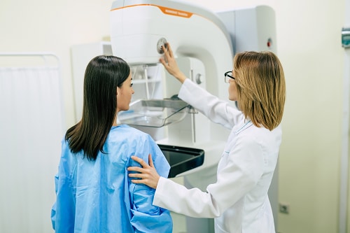
Radiation in Healthcare: Mammogram
Mammograms for breast cancer screening can help find breast cancer early—before there are any signs or symptoms. A mammogram, or mammography exam, uses x-ray beams (a form of ionizing radiation) to create images of the inside of the breast. Healthcare providers use mammograms to check for breast cancer and other conditions that may cause symptoms in the breast of both men and women.
Learn more about mammograms and breast cancer.
Finding and treating cancer early can greatly improve the chances of full recovery and may mean shorter and less invasive treatments. Some risks from mammograms include false results and exposure to a small dose of ionizing radiation during the procedure. We all are exposed to ionizing radiation every day from the natural environment, but additional exposures can lead to an increase in the possibility of developing cancer later in life.
What You Should Know

Your healthcare provider may recommend a mammogram for breast cancer screening or for other symptoms in the breast like a lump, pain, changes in breast skin texture (such as turning red, dimpled, or puckered), or nipple discharge.
The United States Preventive Services Task Forceexternal icon recommends:
- Mammograms every two years for women who are 50-74 years old and are at average risk for breast cancer.
- Women who are 40-49 years old should talk to their healthcare provider about when or how often to get a mammogram based on personal risk and health history.
Work with your healthcare provider to decide on the best screening process and schedule for you. Your healthcare provider may recommend a mammogram when they believe that benefits outweigh the risks for your health.
What To Expect
Find information on special considerations for pregnant women.
Before The Procedure
- Make sure to let your healthcare provider or radiologist (medical professional specially trained in radiation procedures) know if you are pregnant or think you could be pregnant.
- Do not wear deodorant, talcum powder, or lotion under your arms or on your breast area on the day of the exam.
- The radiologist will tell you what clothing or other items you are wearing (such as jewelry or eyeglasses) to remove so they can get clear images.
During The Procedure
- Your breast will be placed on a special platform and compressed with a clear plastic paddle. You may experience discomfort.
- You will be asked to change positions between images. The routine views are a top-to-bottom view and an angled side view. The process will be repeated for the other breast.
- You must hold very still and may be asked to hold your breath for a few seconds while the x-ray picture is taken to reduce the possibility of a blurred image.
- The information collected will be processed to show the structures in the breast and any abnormalities.
Benefits
Mammograms
- Are specifically designed to look for breast diseases and can find cancers up to three years before they can be felt. Early detection helps successful treatment.
- Can track changes in breast tissue over time.
- Help guide treatment when cancer is present.
Risks
- Results can sometimes be false.
- A false-positive test, one that indicates you may have cancer when you truly do not, may lead to extra expenses, testing, and stress.
- A false-negative test, one that says you do not have any cancer when you truly do, may delay your diagnosis and treatment.
If your doctor suspects your results could be false, he or she may repeat testing or order a different type of test like an ultrasound. This is especially common in those who have dense breasts.
Related Links
American College of Radiology
FDA
Image Gently
- What Parents should Know about Medical Radiation Safety pdf icon[PDF – 430kb]external icon
- Educational Materialsexternal icon
EPA
- RadTown USA Medical X-Raysexternal icon
- Radiation Protection Guidance for Diagnostic and Interventional X-Ray Proceduresexternal icon
National Library of Medicine