Key points
- Up to 225 cases of leprosy are identified in the U.S. every year.
- There are three types of leprosy, each with slightly different signs and symptoms.
- People in the U.S. with the disease may be treated at specialized clinics run by the National Hansen's Disease Program.
- Leprosy can be cured with a combination of antibiotics.

Etiology
Leprosy (Hansen's disease) is caused by acid-fast rod-shaped bacillus Mycobacterium leprae and Mycobacterium lepromatosis. The organism multiplies very slowly (dividing approximately once every 13 days).
It is an obligate intracellular pathogen that lacks several genes needed for independent survival. Thus, it has never been grown in bacteriologic media. However, it has been grown in mouse footpads.
Who is at risk
Leprosy is very rare in the United States, with up to 225 cases reported per year. Most people with Hansen's disease in the U.S. became infected overseas in a country where it is prevalent.
There are cases of leprosy reported in U.S. residents with no international travel. Several cases in the southern United States were likely contracted from armadillos.
Clinical features
Leprosy has a range of clinical manifestations based on the immune response to the bacteria. There are two main systems of classification, the Ridley-Jopling and the World Health Organization (WHO) classification system. The five-group Ridley-Jopling classification covers the clinical spectrum of leprosy while the simplified two-group WHO classification, including paucibacillary (PB) and multibacillary (MB) leprosy, is used for treatment purposes.
Based upon signs and symptoms and histopathological findings, the Ridley-Jopling system classifies leprosy from a spectrum of high cell-mediated immunity to low cell-mediated immunity including tuberculoid (TT), borderline tuberculoid (BT), mid-borderline (BB), borderline lepromatous (BL), and lepromatous (LL).
Tuberculoid (TT) leprosy
This is characterized by limited disease with the presence of a few well-defined hypopigmented skin lesions with marked sensory loss. This is due to infection of the peripheral nerves supplying the region. The body's immune response may also result in swelling of the peripheral nerves. In the absence of treatment, the lesions enlarge slowly. This type of leprosy may be self-healing.
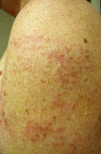
Borderline Tuberculoid (BT) leprosy
When cellular immunity is high but not as strong as TT, skin lesions look like TT lesions, but there are more than 6 lesions with very few bacilli.
Mid-borderline (BB) leprosy
In BB, the lesions show a mixed appearance. Usually, BB lesions have very clearly defined areas of central healing with somewhat less-defined outer edges.
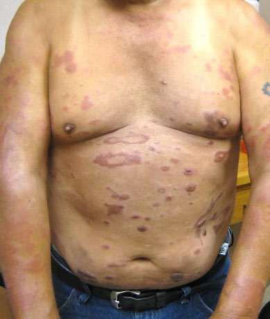
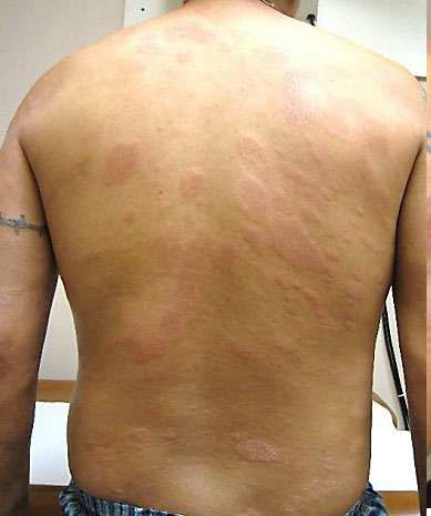
Borderline Lepromatous (BL) leprosy
When cellular immunity is lower, skin lesions look more like lepromatous (LL) lesions, but the macules and nodules are more sharply defined. Lesions are asymmetrical, and there is normal skin between lesions.
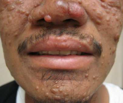
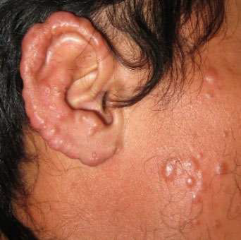
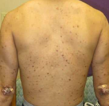
Lepromatous (LL) leprosy
LL leprosy represents the extreme end of the spectrum where the patient has essentially no cell-mediated immunity to the infection and the bacilli multiply uncontrollably. The highest concentrations of bacilli are found in the skin and nerves. The skin lesions are numerous and may have vague margins. There is usually little or no loss of sensation in the skin in the early lesions. There is generalized infiltration of the skin, usually in the cooler zones of the body such as the extremities or eyebrows.
Other presentation
Primary neural leprosy (PNL)
PNL is characterized by isolated involvement of peripheral nerve trunks with no skin lesions. It is more common in endemic countries than in the U.S. Diagnosis of PNL is extremely difficult; it's commonly diagnosed without histologic examination of tissue biopsies.
Testing and diagnosis
Leprosy is diagnosed based on clinical presentation, and the diagnosis is confirmed by skin or nerve biopsy and acid-fast staining.
Treatment and recovery
Treat leprosy using a combination of anti-leprosy drugs. There are multiple treatment options and protocols available.
The major anti-leprosy drugs include rifampin, moxifloxacin, minocycline, dapsone, clarithromycin, and clofazimine. WHO recommends multi-drug therapy of dapsone, rifampicin, and clofazimine with different lengths of treatment based on the type of disease.
Once treatment begins, the patient is no longer considered infectious. Treatment usually lasts 1 to 2 years. Leprosy can be cured if treatment is completed as prescribed.
Reactions to treatment
Most leprosy patients will experience an acute hypersensitivity or immunological reaction to the M. leprae organism during their disease. Reactions may occur before treatment begins or after it is completed. Reactions are not due to medications used to treat the disease, although people treated with clofazimine tend to have slightly fewer episodes.
There are two main groups of reactions:
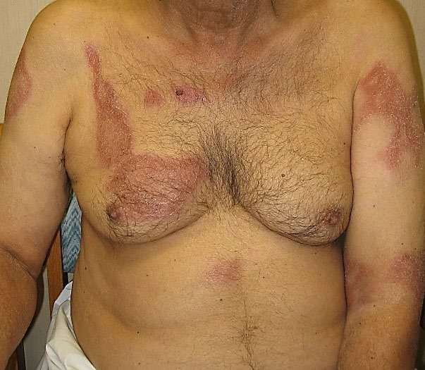
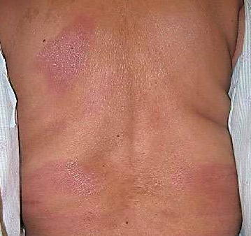
- Type 1 reactions, also called reversal reactions, are typical in borderline leprosy (BT, BB, BL). They show up as edema and erythema of pre-existing lesions. In some cases, neuritis and new lesions or fever may occur, though it's rare.
- Type 2 reactions, or Erythema Nodosum Leprosum (ENL), are most frequently seen in patients with MB leprosy. Patients usually present with painful erythematous nodules often distributed between existing lesions and moderate-to-high fever. Inflammation of other tissues may be present, including peripheral neuritis, orchitis, lymphadenitis, iridocyclitis, nephritis, periostitis and arthralgias.
Lucio's phenomenon is a rare and severe necrotizing reaction typically seen in patients of Mexican ancestry with MB leprosy.
Long-term effects
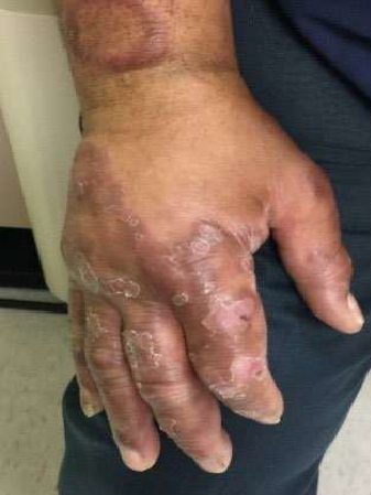
Over time, if leprosy is not treated, leprosy can gradually affect nerves and tissues, sometimes causing structural changes to the nose and hands. Early diagnosis and treatment, before nerve damage occurs is the best prevention of deformity and disability. Routine care for insensitive eyes, hands, and feet is crucial to minimize and prevent deformities.
To prevent complications, the NHDP has developed videos for providers on performing the eye, hand, and foot screens in patients with leprosy: