Key points
- Skin snips are the most common ways of diagnosing onchocerciasis.
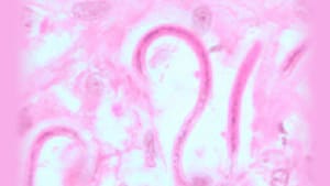
- A healthcare professional will example your skin under a microscope to look for the microfilariae or immature worms.
- You can also have your eyes examined for signs of damage caused by the infection.

Testing methods
If a healthcare provider thinks you might have onchocerciasis, there are several ways to confirm the diagnosis. They can
- Examine a skin snip—a thin skin biopsy—to look for the parasite microfilariae through a microscope. Typically, your provider will collect six snips from different areas of the body. This is the most common method of diagnosis.
- Surgically remove a nodule (lump) from your skin and examine it for adult worms.
- Examine your eye for signs of damage caused by microfilariae, or for the adult worms.
- Give you a blood test to look for signs of antibodies your immune system produced in response to an infection. However, antibody tests can’t distinguish between past and current infections. They are not as useful in people who lived in areas where the parasite is spread, but they are useful in visitors to these areas. Some antibody tests are general tests for infection with any filarial worm, and some are more specific for onchocerciasis.
Diagnosing onchocerciasis can be difficult in light infections (little or low presence of the parasite). This is more common in people who have traveled to but don't live in affected areas.
