At a glance
- Patients infected with O. volvulus present with a range of symptoms from none to skin rashes (usually itchy), eye disease, or nodules under the skin.
- Microscopic examination for microfilariae using skin snips is the best way to diagnose onchocerciasis.
- Antibody tests can identify filarial disease, but not specific diseases.
- There are no vaccines or medications available to prevent onchocerciasis.
Cause
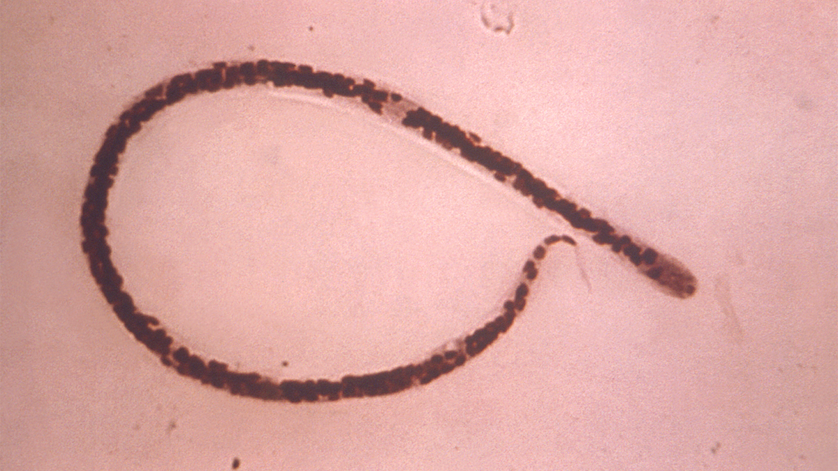
Onchocerciasis is caused by the filarial worm Onchocerca volvulus and spreads through repeated bites by blackflies of the genus Simulium. When a blackfly bites a person with onchocerciasis, the blackfly consumes microscopic immature worms, or microfilariae, from the infected person's skin. The microfilaria develop into larvae, which overtime become infectious to people. When the blackfly is biting a human, the larvae leave the fly. The larvae then penetrate the skin to infect the person. The intensity of human infection (number of worms in an individual) is related to the number of infectious bites sustained by an individual.
Clinical features
Most people with onchocerciasis have no symptoms. However, people with heavy infections will usually have one or more of three conditions: skin rash (usually itchy), eye disease, or nodules under the skin. Additionally, blindness is usually seen in the setting of longstanding and intense infection.
Prevention
There are no vaccines or medications available to prevent becoming infected with O. volvulus. The best prevention efforts include personal protection measures against biting insects. CDC provides guidance and resources on preventing mosquito bites while traveling that you can share with patients. Although onchocerciasis is transmitted by blackflies and not mosquitoes, guidance for preventing bites is similar.
Testing and diagnosis
The test for diagnosing onchocerciasis remains the skin snip biopsy. The biopsy is performed using a sclerocorneal biopsy punch or by elevating a small cone of skin (3 mm in diameter) with a needle and shaving it off with a scalpel. This will result in the removal of around 2 mg of tissue.
The tissue is then incubated in normal saline at room temperature for 24 hours to allow the microfilariae (larvae) to emerge. The microfilariae can then be identified microscopically. The sites for the skin snip are usually over the iliac crest, the scapula, and the lower extremities. Six snips provide the most diagnostic sensitivity. Skin snip sensitivity may be limited in the pre-patent stage of infection, which can last approximately 12 – 18 months, and in low intensity infections. Performing polymerase chain reaction (PCR) of the skin snip can increase the sensitivity of skin snip for low intensity infections though this is not commercially available.
If a patient has skin nodules caused by Onchocerca infection, nodulectomy allows for the identification of adult worms (macrofilariae) in the tissue. Use slit lamp eye exams to visualize microfilariae, or the lesions they cause, in individuals with eye disease.
Antibody tests
There are antibody tests that can assist in the diagnosis of onchocerciasis, though many are not available outside the research setting. There is a general screen for any filarial infection (including Wuchereria, Brugia, Loa, and Mansonella infections) that is available in some specialty diagnostic labs. Because the test is highly sensitive, it is useful in determining if someone has had a filarial infection, but it is not specific enough to identify which filarial infection. There are several Onchocerca-specific serologic tests in existence, such as the OV-16 antigen antibody test and the OV luciferase immunoprecipitation system (LIPS) assay, but these are currently only available in research settings and are not approved for diagnosis in the United States.
As with any antibody test, the results indicate only that the patient has been exposed to a filarial disease. It cannot determine if the patient has an active infection. This distinction is less important in symptomatic travelers, but it limits the usefulness of the test in people from endemic areas.
One advantage of the test is that it can pick up evidence of infection in the pre-patent stage of infection. In general, the diagnosis of O. volvulus infection should be made with skin snip. However, when skin snips are negative and clinical suspicion of infection is high, the general antibody test could exclude infection.
If a general antibody test is positive, it might be necessary to perform additional skin snips and/or seek additional diagnostic information by enlisting the assistance of researchers who perform additional antibody tests.