Volume 29, Number 8—August 2023
Dispatch
Emerging Corynebacterium diphtheriae Species Complex Infections, Réunion Island, France, 2015–2020
Abstract
Clinical, epidemiologic, and microbiologic analyses revealed emergence of 26 cases of Corynebacterium diphtheriae species complex infections on Réunion Island, France, during 2015–2020. Isolates were genetically diverse, indicating circulation and local transmission of several diphtheria sublineages. Clinicians should remain aware of the risk for diphtheria and improve diagnostic methods and patient management.
Diphtheria is a contagious, potentially fatal infection caused by toxin-producing bacteria of the Corynebacterium diphtheriae species complex, which includes C. diphtheriae, C. ulcerans, C. pseudotuberculosis, C. rouxii, C. belfantii, and C. silvaticum. Infection is localized principally in the upper respiratory tract, and production of diphtheria toxin (encoded by the tox gene) can cause systemic complications. Cutaneous diphtheria and diphtheria endocarditis can also act as sources of respiratory infections (1–4). Diphtheria surveillance has traditionally focused on respiratory illness caused by toxigenic C. diphtheriae but has been expanded in some countries to include all C. diphtheriae species complex infections irrespective of species, infection site, or toxigenicity, enabling broader disease monitoring. C. diphtheriae spreads via human-to-human contact; C. ulcerans and C. pseudotuberculosis are transmitted to humans primarily through animal contact.
Diphtheria was once a major cause of infant death, but global incidence has declined over the past century, largely because of mass vaccination. Consequently, diphtheria is now often considered a forgotten disease (5). Nevertheless, diphtheria reemergence has been reported in high-income countries and is closely related to patient travel history. Diphtheria is considered endemic in Madagascar, Comoros, and Mayotte in the southwest Indian Ocean, but few cases have been reported on other islands, including Réunion Island, an overseas department of France, where cases emerged in 2015 (6,7). Vaccination coverage is poorer in Mayotte (45% for 7- to 11-year-old children) than in Réunion Island (96% for children 11 months of age). Recent improvements in laboratory diagnostic capabilities, such as mass spectrometry use, have increased reports of C. diphtheriae species complex infections (8). However, knowledge of prevalence and origin of those infections is limited in this region. The aims of this study were to review the clinical, epidemiologic, and microbiologic characteristics of C. diphtheriae species complex infections on Réunion Island during 2015–2020 and identify possible links with cases on other islands in the region.
We included all cases of C. diphtheriae species complex infections reported to the regional health agency and recorded at Réunion Island University Hospital during 2015–2020. We analyzed medical records and extracted age, sex, country of residence, recent travel, contact with animals, socioeconomic status, and diphtheria vaccination status for each case. We performed antimicrobial susceptibility testing; identified co-infecting strains; and determined tox gene presence, diphtheria toxin production, and biovar and sequence type (ST). We sent each isolate to the National Reference Center for Corynebacteria of the diphtheriae Complex (Institut Pasteur, Paris, France) to confirm species identity through multiplex PCR and biotyping as previously described (8–10). We detected the tox gene by using conventional PCR or, since 2019, by using multiplex real-time PCR (10). We assessed toxin production by using a modified Elek test (11). We determined antimicrobial drug susceptibility by using disk diffusion or by determining MICs (E-test; bioMérieux, https://www.biomerieux.com), in accordance with CASFM/EUCAST2021 (https://www.sfm-microbiologie.org/2021/04/23/casfm-avril-2021-v1-0) recommendations for benzylpenicillin, amoxicillin, cefotaxime, clindamycin, rifampin, and ciprofloxacin. We genotyped each isolate by using multilocus sequence typing (MLST) (12).
Figure
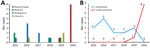
Figure. Number of cases diagnosed per year in study of emerging Corynebacterium diphtheriaespecies complex infections, Réunion Island, France, 2015–2020. Number of cases were classified according to geographic origin (A)...
A total of 26 cases of C. diphtheriae species complex infections were recorded, from which 27 C. diphtheriae and 2 C. ulcerans isolates were cultured. Most (88.5%) infected patients were male; median age was 60 (interquartile range 32.5–67) years. Fourteen (50%) patients lived on Réunion Island, 3 (11.5%) in Mayotte, 4 (19.2%) in mainland France, 3 (11.5%) in Comoros, and 2 (7.8%) in Madagascar. Most (84.6%) patients had skin manifestations, and 16 patients were vaccinated (Table 1; Appendix Figure). Of 24 C. diphtheriae infections, 8 occurred in patients who had recently traveled to or originated from Madagascar, 4 who traveled to or originated from Mayotte, and 3 who traveled to or originated from Comoros. Since 2018, a total of 9 cases on Réunion Island have been considered locally acquired; all of those patients lived in poor socioeconomic conditions. C. ulcerans infections occurred in 2 patients living on Réunion Island who had not traveled recently but had contact with animals (Table 1; Figure). We performed a Spearman rank correlation to compare locally acquired strains isolated during 2015–2018 and 2019–2020; a 75% increase in locally acquired C. diphtheriae infections occurred in 2019–2020 (ρ = 0.8452; p = 0.0341).
Isolates were obtained from cutaneous lesion (n = 24), bone (n = 4), and respiratory (n = 1) samples. Eight of 27 C. diphtheriae isolates were toxigenic, yielding positive Elek test results. The 2 C. ulcerans isolates were nontoxigenic. C. diphtheriae isolates were characterized as biovars Mitis (n = 20) and Gravis (n = 7).
Patient isolates were co-infected most frequently with Staphylococcus aureus (n = 17) and Streptococcus pyogenes (n = 18). Benzylpenicillin resistance was observed in 80% of isolates according to CASFM/EUCAST2021 recommendations, but isolates were categorized as susceptible increased exposure according to EUCAST version 13.0 proposed breakpoints (https://www.eucast.org/clinical_breakpoints) (Appendix Table). One (3.5%) C. diphtheriae isolate was resistant to amoxicillin (CD8/FRC0402; MIC 1.5 mg/L), and 1 was resistant to rifampin. Both C. ulcerans isolates were resistant to clindamycin (100%, natural low susceptibility), whereas clindamycin resistance was observed for only 1 C. diphtheriae isolate.
We identified 21 STs by MLST analysis, including ST88 for C. diphtheriae isolates from 4 patients and ST339 for both C. ulcerans isolates (Table 2). All C. diphtheriae STs had 2–5 mismatches, except ST87 and ST237, which had 1 mismatch between them. ST339 (C. ulcerans) had 7 mismatches with all C. diphtheriae STs.
We report increased prevalence of cutaneous C. diphtheriae species complex infections on Réunion Island during 2015–2020. Introduction of mass spectrometry analysis in hospital laboratories and increased clinician awareness might have led to increased case reporting. Our study confirms that C. diphtheriae species complex members are circulating and are likely underestimated in the southwest Indian Ocean (7,13). Moreover, we observed emergence of locally acquired cutaneous C. diphtheriae infections on Réunion Island since 2019. The number of imported cases in 2020 was probably limited because of the COVID-19 pandemic, which reduced travel. Indeed, all C. diphtheriae cases identified during 2015–2018 occurred in patients who had traveled from other islands in the Indian Ocean. In addition, cutaneous diphtheria appeared to be associated with poor socioeconomic living conditions, in which alcoholism, drug dependence, and homelessness are factors that increase risk for human-to-human transmission and virulence (14).
A total of 8 (30%) C. diphtheriae isolates were toxigenic and caused cutaneous infections. Nontoxigenic C. diphtheriae isolates (70%, n = 19) were obtained from cutaneous lesions, respiratory samples, and bone samples. Clinicians should be aware that nontoxigenic C. diphtheriae can potentially cause severe disease (1,14,15). Moreover, all isolates were co-infected with pyogenic bacteria, suggesting diphtheria infection should be considered under polymicrobial conditions.
MLST analysis identified 21 different STs; most were unrelated (>2 mistmatches) reflecting marked genetic diversity of isolates. ST88 was found in 4 patients living on Réunion Island who had not traveled recently, indicating probable local acquisition. ST88 had previously been reported only in patients from Mayotte. Therefore, our results show that multiple C. diphtheriae species complex clones are circulating in the southwest Indian Ocean (8). Both C. ulcerans strains belonged to ST339. The National Reference Center reported that ST339 is the predominant C. ulcerans ST found in animals in France. Although considerable ST diversity was revealed, whole-genome sequencing will be required to further evaluate circulating C. diphtheriae clones in this region.
In conclusion, we describe emergence of locally acquired C. diphtheriae species complex infections on Réunion Island during 2019–2020. Local clinicians and microbiologists should remain aware of this neglected infection; improvements should be made in diagnostic methods and management of infected patients, such as maintaining availability of diphtheria antitoxin.
Dr. Garrigos is a research scientist in the microbiology department of Félix Guyon University Hospital of Réunion Island, France. His research interests focus on bacterial diseases, antimicrobial resistance, cystic fibrosis patients, and emerging infectious diseases.
Acknowledgment
The National Reference Center for Corynebacteria of the diphtheriae Complex is supported financially by Santé publique France (Saint-Maurice, France).
References
- Patey O, Bimet F, Riegel P, Halioua B, Emond JP, Estrangin E, et al.; Coryne Study Group. Clinical and molecular study of Corynebacterium diphtheriae systemic infections in France. J Clin Microbiol. 1997;35:441–5. DOIPubMedGoogle Scholar
- Hadfield TL, McEvoy P, Polotsky Y, Tzinserling VA, Yakovlev AA. The pathology of diphtheria. J Infect Dis. 2000;181(Suppl 1):S116–20. DOIPubMedGoogle Scholar
- Levi LI, Barbut F, Chopin D, Rondeau P, Lalande V, Jolivet S, et al. Cutaneous diphtheria: three case-reports to discuss determinants of re-emergence in resource-rich settings. Emerg Microbes Infect. 2021;10:2300–2. DOIPubMedGoogle Scholar
- Sangal V, Hoskisson PA. Evolution, epidemiology and diversity of Corynebacterium diphtheriae: New perspectives on an old foe. Infect Genet Evol. 2016;43:364–70. DOIPubMedGoogle Scholar
- Sharma NC, Efstratiou A, Mokrousov I, Mutreja A, Das B, Ramamurthy T. Diphtheria. Nat Rev Dis Primers. 2019;5:81. DOIPubMedGoogle Scholar
- Scheifer C, Rolland-Debord C, Badell E, Reibel F, Aubry A, Perignon A, et al. Re-emergence of Corynebacterium diphtheriae. Med Mal Infect. 2019;49:463–6. DOIPubMedGoogle Scholar
- Belchior E, Henry S, Badell E, Collet L, Benoit-Cattin T, de Montera AM, et al. Diphtheria in Mayotte, 2007-2015. Emerg Infect Dis. 2017;23:1218–20. DOIPubMedGoogle Scholar
- Hennart M, Panunzi LG, Rodrigues C, Gaday Q, Baines SL, Barros-Pinkelnig M, et al. Population genomics and antimicrobial resistance in Corynebacterium diphtheriae. Genome Med. 2020;12:107. DOIPubMedGoogle Scholar
- Dazas M, Badell E, Carmi-Leroy A, Criscuolo A, Brisse S. Taxonomic status of Corynebacterium diphtheriae biovar Belfanti and proposal of Corynebacterium belfantii sp. nov. Int J Syst Evol Microbiol. 2018;68:3826–31. DOIPubMedGoogle Scholar
- Badell E, Guillot S, Tulliez M, Pascal M, Panunzi LG, Rose S, et al. Improved quadruplex real-time PCR assay for the diagnosis of diphtheria. J Med Microbiol. 2019;68:1455–65. DOIPubMedGoogle Scholar
- Engler KH, Glushkevich T, Mazurova IK, George RC, Efstratiou A. A modified Elek test for detection of toxigenic corynebacteria in the diagnostic laboratory. J Clin Microbiol. 1997;35:495–8. DOIPubMedGoogle Scholar
- Bolt F, Cassiday P, Tondella ML, Dezoysa A, Efstratiou A, Sing A, et al. Multilocus sequence typing identifies evidence for recombination and two distinct lineages of Corynebacterium diphtheriae. J Clin Microbiol. 2010;48:4177–85. DOIPubMedGoogle Scholar
- Rakotomalala RS, Andrianirina ZZ, Ratsima E, Randrianandraina P, Randrianirina F, Edosoa GT, et al. Corynebacterium diphtheriae infection in Mahajanga, Madagascar: first case report. J Trop Pediatr. 2021;67:fmaa064.
- Badenschier F, Berger A, Dangel A, Sprenger A, Hobmaier B, Sievers C, et al. Outbreak of imported diphtheria with Corynebacterium diphtheriae among migrants arriving in Germany, 2022. Euro Surveill. 2022;27:
2200849 . DOIPubMedGoogle Scholar - Farfour E, Badell E, Zasada A, Hotzel H, Tomaso H, Guillot S, et al. Characterization and comparison of invasive Corynebacterium diphtheriae isolates from France and Poland. J Clin Microbiol. 2012;50:173–5. DOIPubMedGoogle Scholar
Figure
Tables
Cite This ArticleOriginal Publication Date: June 14, 2023
1These first authors contributed equally to this article.
2These authors contributed equally to this article.
Table of Contents – Volume 29, Number 8—August 2023
| EID Search Options |
|---|
|
|
|
|
|
|
Please use the form below to submit correspondence to the authors or contact them at the following address:
Thomas Garrigos, Laboratoire de Microbiologie, Hôpital Universitaire Félix Guyon, Allée des Topazes, 97400 Saint-Denis, La Réunion, France
Top