Key points
- Nuclear medicine can be used by healthcare providers for both diagnosis and treatment of tissues and organs.
- Some nuclear medicine procedures are longer and use more radiation than others.
- You may give off small amounts of radiation after your procedure and need to take steps to protect others from exposure.

The basics
Nuclear medicine uses radioactive material inside the body for two reasons:
- To see how organs or tissue are functioning (diagnosis).
- To target and destroy damaged or diseased organs or tissue (treatment).
We are all exposed to ionizing radiation every day from the natural environment. However, added exposures like those from nuclear medicine procedures can slightly increase the risk of developing cancer later in life.
Talk to your healthcare provider
When it's used for diagnosis
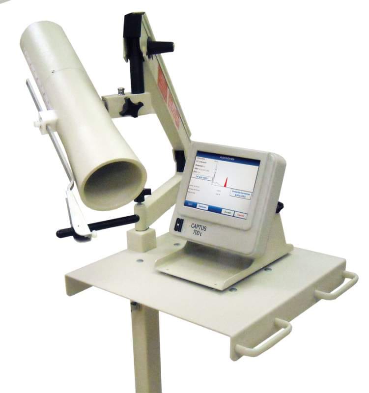
Nuclear medicine can show how the organs or tissues are functioning. For most diagnostic procedures, a tracer, which contains the radioactive material, is injected, swallowed, or inhaled. Then the healthcare provider or radiologist (a healthcare professional with special training to use radiation in healthcare) observes the tracer. They use a radiation detector to see how much of the tracer is absorbed or how it reacts. This will give the provider information about how well organs or tissues are working.
Common uses of nuclear medicine for diagnosis include scans of the heart, lungs, kidneys, gallbladder, and thyroid.
In a type of nuclear medicine called positron emission tomography (PET), the tracer shows the natural activity of cells. This provides more detailed information on how organs are working and if there is damage to the cells. PET scans are often combined with computed tomography (CT) scans or magnetic resonance imaging (MRI). These scans provide three-dimensional images of the organ.
Common uses of PET scans include:
- Diagnosing heart disease, Alzheimer's disease, and brain disorders.
- Getting detailed information about cancerous tumors to decide the best treatment option.
When it's used for treatment
When used in treatment, the tracer targets a harmful organ or tissue and damages or stops the growth of its cells.
Two common uses of nuclear medicine for treatment include radioactive iodine therapy and brachytherapy. Brachytherapy is a form of treatment where a sealed radiation source is placed inside or next to the area requiring treatment.
What to expect
Before the procedure
- You will receive a tracer either through injection, inhalation (breathing it in), or through a pill or substance to swallow.
- You may need to wait a certain amount of time for the tracer to travel through your body to the tissue or organ being diagnosed or treated.
During the procedure
- You may be asked to lie down on a table or to walk on a treadmill.
- A camera that detects radiation will be placed over your body to collect information on how the tracer is acting in an organ or tissue.
After the procedure
- The radiologist and your healthcare provider use this information to see how an organ or tissue is functioning.
- The radioactive material from the tracer will pass out of your body in a few hours to a few days, depending on the type of tracer and test you receive.
When You Go Home
Right after your procedure, your body is very slightly radioactive (giving off radiation). The radiation wears off with time and is not directly harmful to others. Your healthcare provider may give you special instructions to help reduce any small amounts of radiation you give off. Drinking a lot of water may help the radioactive material leave your body quicker.
Before you leave, ask your healthcare provider if there are steps you should take to protect others. You should also discuss any concerns or questions about the information you were given. Talk to your healthcare provider if you are currently breastfeeding.
The ionizing radiation dose for these procedures is typically higher than the dose received from a common x-ray procedure. There are always some possible risks from exposure to ionizing radiation in healthcare, but these procedures should be used when the health benefits outweigh these risks.
Impacts
Benefits
- Provides information on how organs, tissues, and cells are working. (Other common imaging procedures only show the structures.)
- Can be used also in targeted treatments to kill or damage harmful or cancerous cells, reduce the size of tumors, or reduce pain.
Risks
- Radiation doses are usually higher than in common imaging like x-rays. This means these procedures are slightly more likely to increase the possibility you may get cancer later in life.
- Some nuclear medicine procedures are longer and use more radiation than others. These could cause skin reddening and hair loss.
- You may give off small amounts of radiation right after your procedure and need to take steps to protect others from exposure.