At a glance
DEXA (dual x-ray absorptiometry) scans measure bone density (thickness and strength of bones) by passing a high- and low-energy x-ray beam (a form of ionizing radiation) through the body, usually in the hip and the spine. DEXA scans are often used to diagnose specific conditions, such as bone thinning.

The basics
DEXA (dual x-ray absorptiometry) is important for diagnosing (seeing if someone has) osteoporosis or bone thinning.
The amount of radiation used in DEXA scans is very low and similar to the amount of radiation used in x-rays. We are all exposed to ionizing radiation every day from the natural environment. However, added exposures can slightly increase the risk of developing cancer later in life.
What you should know
Your healthcare provider may recommend a DEXA scan to test for osteoporosis or thinning of your bones.
Screening for osteoporosis is recommended for women who are 65 years old or older and for women who are 50 to 64 and have certain risk factors, such as having a parent who has broken a hip. However, there are other risk factors for osteoporosis besides age and sex, such as some intestinal disorders, multiple sclerosis, or low body weight. Your healthcare provider may recommend a DEXA scan if you have any of these other risk factors.
DEXA scans should be used when the health benefits outweigh the risks. Talk to your healthcare provider about any concerns you have before a DEXA scan.
What to expect
Before the procedure:
- Make sure to let your healthcare provider or radiologist (a medical professional specially trained in radiation procedures) if you are pregnant or think you may or could be pregnant.
- Dress in loose, comfortable clothing. Don't wear anything that has metal on it like buckles, buttons, or zippers. Metal can interfere with test results.
Find information on special considerations pregnant women and children.
During the procedure:
- You may be asked to remove jewelry, eyeglasses, and any clothing that may interfere with the imaging.
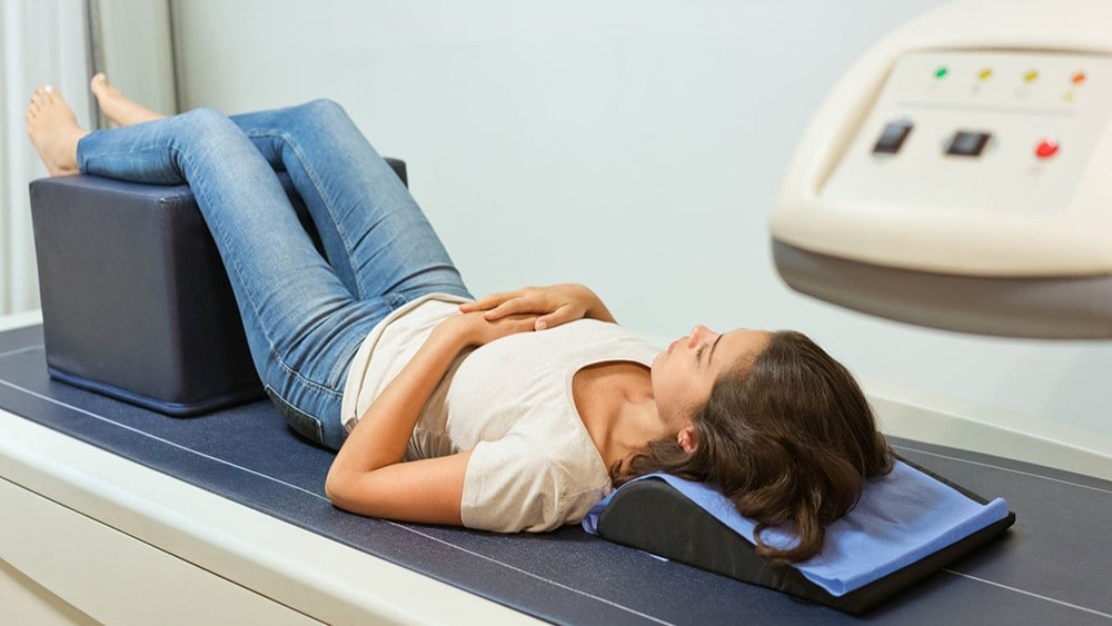
- You will lay on a table and the radiologist or medical assistant will position your legs on a padded box. They also may place your foot in a device so that your hip is turned inward.
- While the image is taken, lay still and follow instructions. You may need to hold your breath for a few seconds.
After the procedure:
- The procedure typically lasts about 15-20 minutes.
- Your healthcare provider will follow up with you with your results. They will show a T-score and a Z-score:
- The T-score shows how your bone density compares to the optimal peak bone density for your sex.
- The Z-score shows how your bone density compares to the bone densities of others who are the same age, sex, and ethnicity.
- The T-score shows how your bone density compares to the optimal peak bone density for your sex.
Impacts
DEXA scans are different from other imaging procedures because they are used to screen for a specific condition.
Benefits:
- Detects weak or brittle bones to help predict the odds of a future fracture.
- Determines if bone density is improving, worsening, or staying the same.
- Can help you and your healthcare provider come up with plans to improve your bone strength and prevent worsening conditions.
Risks:
- A very slight increase in possibility of future cancer, similar to the risks from x-rays.
Resources
American College of Radiology (ACR)
American Society of Radiologic Technologists (ASRT)
Image Gently
U.S. Environmental Protection Agency (EPA)
- RadTown USA Medical X-Rays
- Radiation Protection Guidance for Diagnostic and Interventional X-Ray Procedure
U.S. National Library of Medicine
