
Philophthalmiasis
[Philophthalmus spp.]
Causal Agents
Zoonotic trematodes in the genus Philophthalmus, also called “eye flukes”, have infected humans on very rare occasions. The three Philophthalmus species conclusively identified in humans are P. lacrymosus (also spelled P. lachrymosus and P. lacrimosus), P. gralli, and P. palpebrarum. Note that in most previously reported cases, species could not be ascertained.
Life Cycle
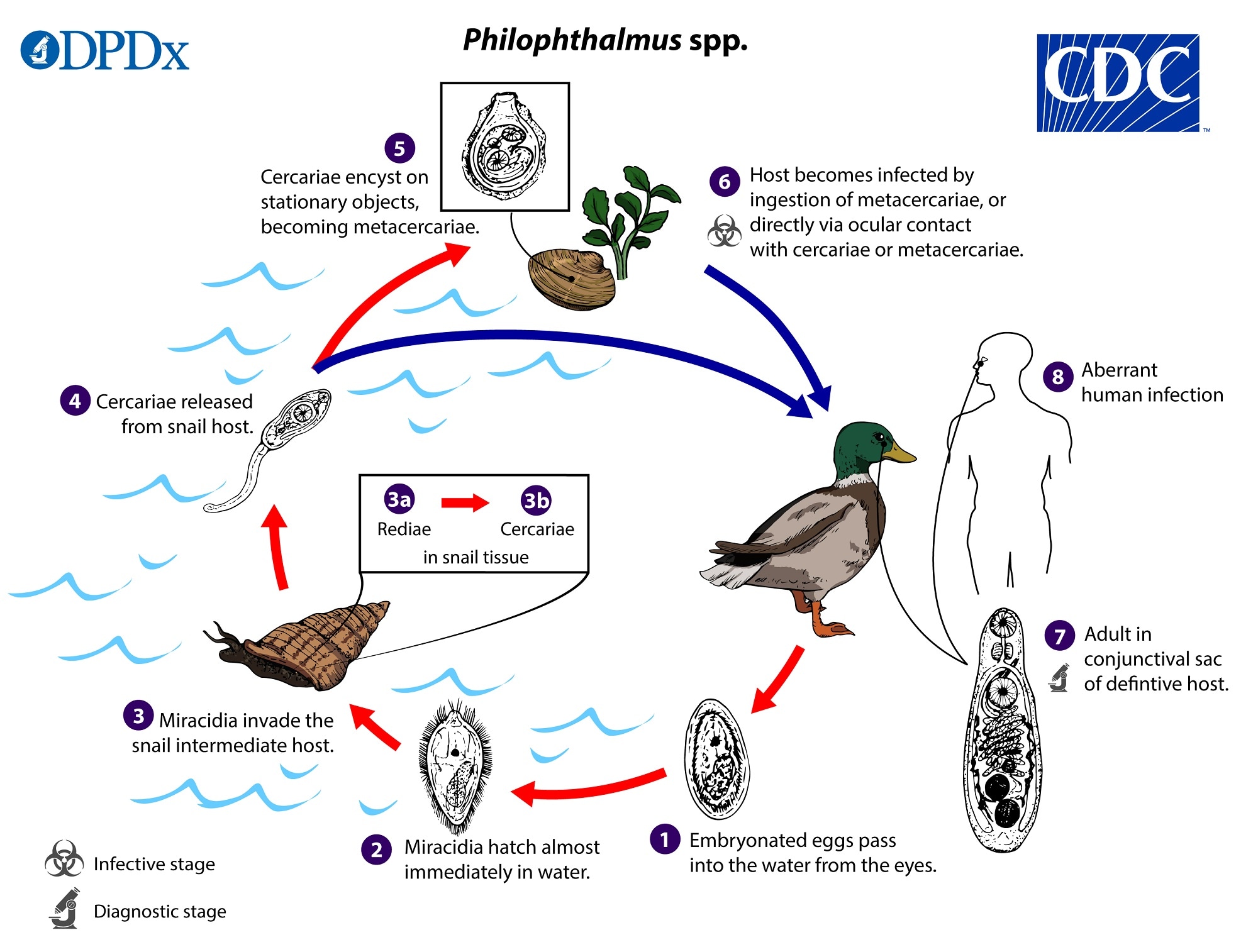
 . Miracidia hatch almost immediately in water
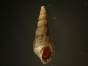
  and penetrate the snail intermediate host
  . Inside the snail host, pre-formed sporocysts within the miracidia produce generations of rediae, which become cercariae (
  ,
,  ). Cercariae are released from the snail
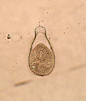
  and encyst on stationary objects in water (e.g. vegetation, shells of mollusks and crustaceans) and become flask-shaped metacercariae; no second intermediate host is involved, as in many other trematodes
  . The definitive host, which is usually an aquatic bird, becomes infected either upon ingestion of metacercariae or by direct exposure of the eye to cercariae or metacercariae in water
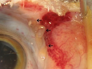
  . When ingested, metacercariae excyst almost instantly in the mouth and migrate through the nasal passages to the ocular orbit over few days. Adult flukes are found in the conjunctival sac, either around the lachrymal glands or under/on the surface of the nictitating membrane
  . Human infections are extremely rare, and the route of infection is not entirely clear. Direct entry of cercariae or metacercariae into the orbit following exposure during bathing or swimming in fresh water seems likely based on some case histories, although oral inoculation cannot be ruled out
  .
.Hosts
The majority of Philophthalmus species are associated with waterbirds and wading birds (e.g., gulls, ducks, geese, herons) although other avian species are capable of being infected under experimental conditions. Aberrant infections have also been achieved experimentally or observed naturally among some mammals, such as rabbits, rats, and non-human primates. All species that have been identified in human infections (P. lacrymosus, P. gralli, P. palpebrarum) use avian hosts.
Snail intermediate hosts are diverse and include freshwater and marine species. Introduced, invasive snail species may be responsible for the spread of Philophthalmus spp. to new locations. For many species, the snail host range is not known. Melanoides tuberculata appears to be the primary intermediate host for P. gralli.
Geographic Distribution
Various Philophthalmus species occur worldwide in avian hosts. Human cases have been reported from North America, Central Europe, the Middle East, East Asia, and South Asia.
Clinical Presentation
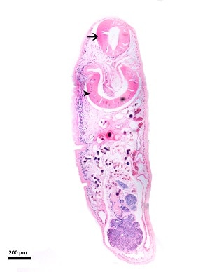
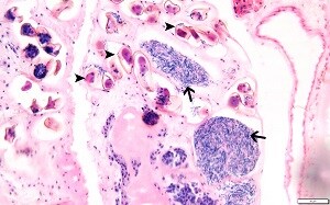
The clinical course of philophthalmiasis in humans has not been well described due to its rarity. The presence of adult flukes in the orbit causes conjunctivitis, keratitis, watery discharge, and mild edema.
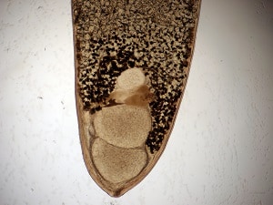
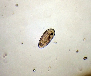
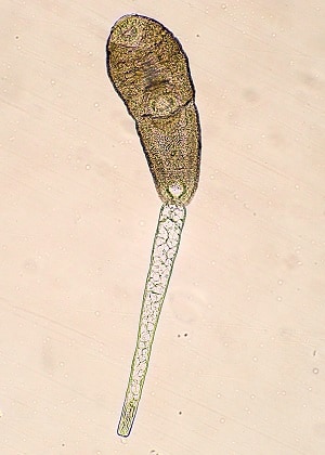
Philophthalmus spp. are small, somewhat elongate trematodes, usually measuring from 2–5 mm in length and 1.5 mm in maximum width (varying by species). Major features are large, muscular oral and ventral suckers, large pharynx, and a long, prominent cirrus pouch. The tegument is generally smooth in appearance although minute spines may be present.
The photographs in this section were provided courtesy of Dr. Hudson Alves Pinto (Department of Parasitology, Universidade Federal de Minas Gerais, Brazil).
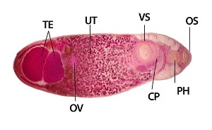
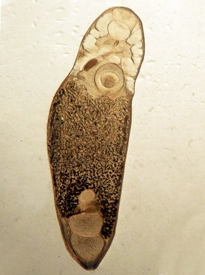
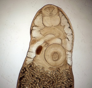
The photographs in this section were provided courtesy of Dr. Hudson Alves Pinto (Department of Parasitology, Universidade Federal de Minas Gerais, Brazil).
Laboratory Diagnosis
Diagnosis is based on the recovery and identification of adult flukes from the ocular orbit. Genus/species identification requires expertise in trematode morphology.
Laboratory Safety
Universal laboratory precautions apply, as only adult fluke specimens are likely to be examined in a diagnostic setting.
Suggested Reading
Waikagul, J., Dekumyoy, P., Yoonuan, T. and Praevanit, R., 2006. Conjunctiva philophthalmosis: a case report in Thailand. The American Journal of Tropical Medicine and Hygiene, 74 (5), pp.848–849.
Lamothe-Argumedo, R., Diaz-Camacho, S.P. and Nawa, Y., 2003. The first human case in Mexico of conjunctivitis caused by the avian parasite, Philophthalmus lacrimosus. Journal of Parasitology, 89 (1), pp.183–185.
DPDx is an educational resource designed for health professionals and laboratory scientists. For an overview including prevention, control, and treatment visit www.cdc.gov/parasites/.